

Early Brain Connections Offer Clues to Later Learning Skills in Very Preterm Infants
Research By: Lili He, PhD | Nehal Parikh, DO, MS
Post Date: November 7, 2026 | Publish Date: Nov. 2, 2026

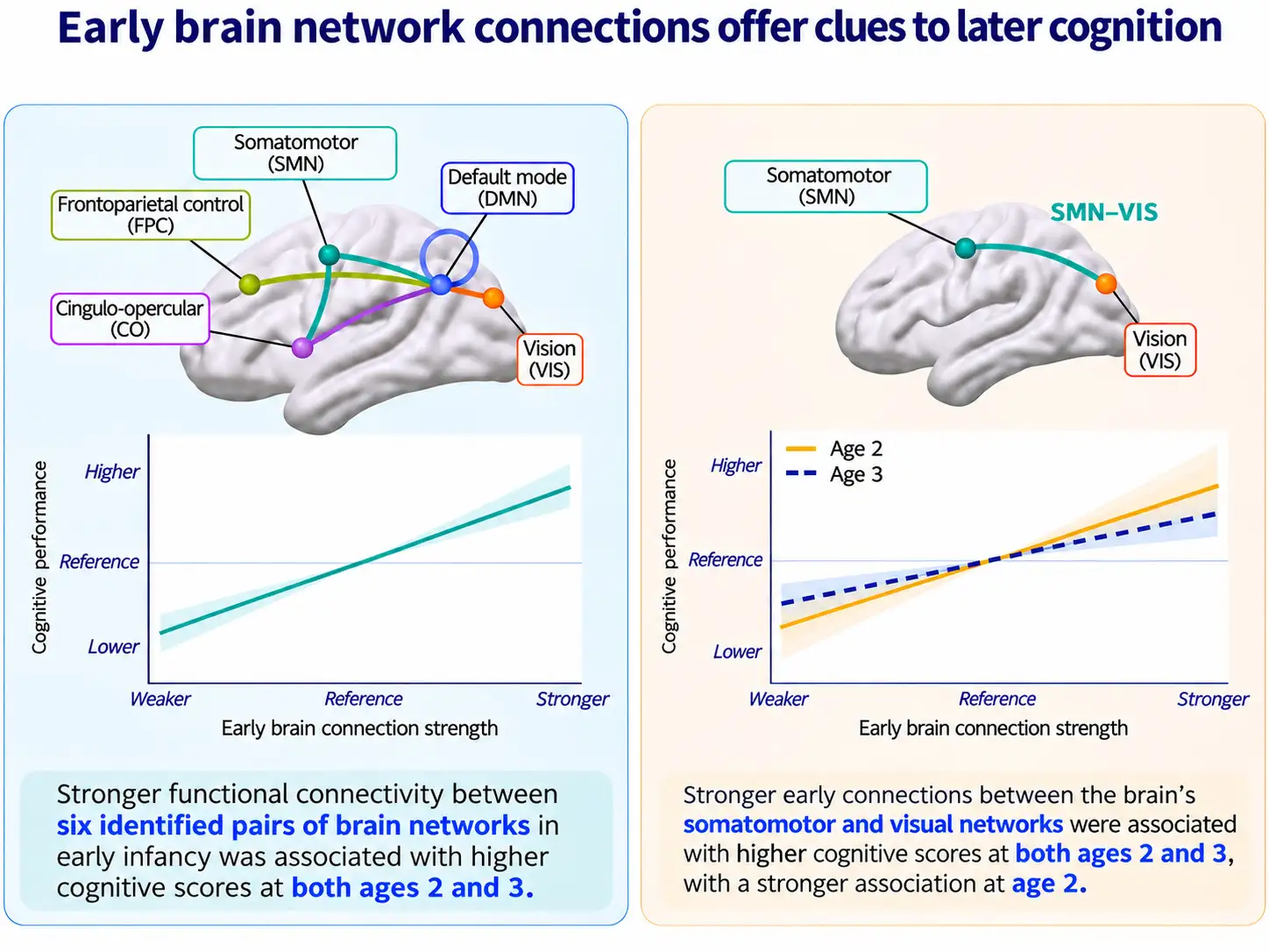
Functional MRI scans of 257 children link newborn brain-network organization with cognitive performance at ages 2 and 3, suggesting a possible new biomarker to guide earlier intervention
For families of babies born very preterm (before 32 weeks gestation), it typically takes several years to discover how much impact their early birth had upon brain development and learning abilities. Now, a study led by experts at Cincinnati Children’s reports that functional MRI scans may serve as an important predictive tool.
In a study of 257 children born very preterm, researchers found that stronger connections involving certain brain networks were associated with better cognitive performance at ages 2 and 3. The findings, published online Nov. 2, 2026, in JAMA Network Open, highlight the potential of neonatal brain imaging to help detect and potentially intervene sooner when developmental difficulties emerge.
The new findings come from the ongoing Cincinnati Infant Neurodevelopmental Early Prediction Study, led by senior authors Nehal Parikh, DO, MS and Lili He, PhD. Junqi Wang, PhD, was first author. Contributing authors included Wei Jia, PhD, Hailong Li, PhD, Zhixiu Lu, PhD, Mekibib Altaye, PhD, Kim Cecil, PhD, and Leanne Tamm, PhD.
“Very preterm infants face an increased risk of cognitive and developmental difficulties, some of which may not become apparent until school age,” Parikh says. “Our study found that patterns of communication between brain regions, measured using functional MRI around the infants’ original due dates, were associated with cognitive performance at ages 2 and 3.”
The findings describe associations that stronger functional brain network connections lead to better cognitive outcomes. It will require further research to determine if and how MRI scans might be used to predict an individual child’s learning needs.
Looking beyond individual brain regions
The researchers used resting-state functional MRI to examine connectivity within and between six brain networks, then evaluated how those early patterns related to cognitive assessments at ages 2 and 3 (adjusted to account for their early births). The analysis accounted for repeated cognitive assessments of the same children, similarities among twins and other multiples, and other factors that could influence cognitive development.
One of the clearest patterns involved the default mode network, or DMN, which participates in internally directed thinking. Stronger connections within this network—and between it and other developing brain systems—were consistently associated with better cognitive performance.
That broader pattern matters.
“The study points not only to how a single network is organized, but also to how networks connect with one another,” He says. “These newborn connections represent an early stage of brain organization, not a fully mature system. This in turn suggests the potential for early intervention to improve outcomes.”
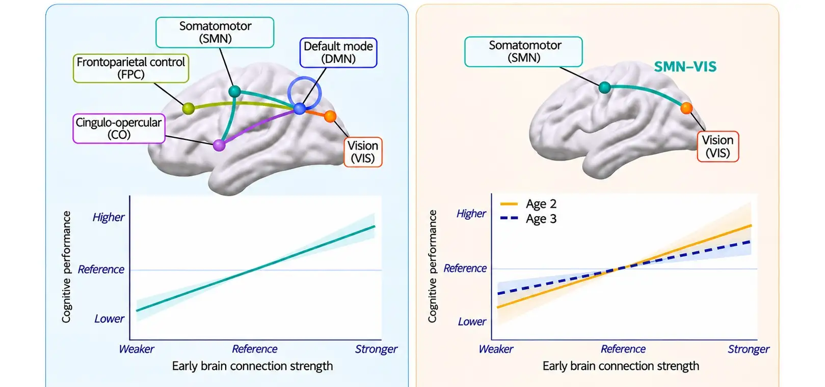
Positive associations also involved the somatomotor network, or SMN, an early-developing system related to movement and sensation. In particular, the SMN’s connections with the visual network and a higher-order functional network called the cingulo-opercularnetwork (CO) were associated with cognitive performance.
A changing relationship as children grow
Not every connection showed the same relationship at both ages.
“The connections associated with cognitive scores were not exactly the same at ages 2 and 3,” Parikh explains. “For example, connections involving regions important for movement and vision showed different relationships at the two ages, suggesting that the link between early brain organization and later cognition may evolve as children grow.”
MRI-based ‘biomarkers’ ahead?
Better understanding of the timing of key moments in brain development eventually could help clinicians, families and educators focus on when to most effectively intervene in the process.
The new findings, by themselves, do not establish a method for using MRI to predict a child’s brain development outcomes. Initially, such scans are more likely to be used by other scientists as a research marker of brain development.
“Additional research needed to develop a validated, stand-alone clinical test is underway to facilitate early diagnosis of learning problems,” Parikh says.
That research is worth pursuing, the co-authors say. Earlier detection could lead to more effective interventions during the critically important first five years of brain development that would allow more children born prematurely to reach their full potential.
Don’t Miss a Post
- Subscribe to the Research Horizons Newsletter
- Follow Cincinnati Children’s Research Foundation on Bluesky, X and LinkedIn
| Original title: | Functional Connectivity at Birth and Longitudinal Neurocognitive Development in Very Pr |
| Published in: | JAMA Network Open |
| Publish date: | Nov. 2, 2026 |
Research By


Latest Posts

About this blog
The Research Horizons blog features news and insights about the latest discoveries and innovations developed by the scientists of Cincinnati Children's. This blog does not provide medical advice, diagnosis, or treatment. Email researchnews@cchmc.org with questions or ideas.
Subscribe to Our Newsletter