

Study Reveals Blood Markers Tied to Eosinophilic Gastritis Disease Activity
Research By: Tetsuo Shoda, MD, PhD
Post Date: October 22, 2026 | Publish Date: October 7, 2026

A Cincinnati Children’s-led study offers a tissue-informed approach to developing future blood biomarkers for eosinophilic gastritis
Key Takeaways
- Researchers identified a blood-based protein signature associated with active eosinophilic gastritis (EoG).
- The signature reflected molecular changes occurring in the stomach tissue of patients with EoG.
- Several blood proteins and inflammatory markers changed as patients achieved remission.
- Findings suggest blood-based tools could one day complement biopsy-based assessments of disease activity.
For people living with eosinophilic gastritis (EoG), keeping track of disease activity often requires repeated endoscopies and tissue biopsies. While these procedures provide valuable information, they can be invasive and may not fully capture the molecular changes occurring within the stomach.
A new study published on October 7, 2026 in The Journal of Allergy and Clinical Immunology found that protein changes occurring in the stomach could also be detected in the blood. The findings provide early evidence that blood-based measures may help researchers better understand EoG activity and response to treatment in the future.
“What is important about this approach is that we started with the diseased tissue and asked which molecular signals could also be seen in the circulation,” says Tetsuo Shoda, MD, PhD, corresponding author of the study. “That gives us a biologically informed path toward developing blood-based tools for monitoring disease.”
A Need for Better Monitoring
EoG is a chronic inflammatory disease characterized by an accumulation of eosinophils, a type of immune cell, in the stomach lining. Monitoring the condition can be challenging because eosinophils are naturally present in the stomach, making it difficult to rely solely on cell counts to understand disease activity.
Previous studies have identified molecular changes in stomach tissue and circulating biomarkers associated with EoG. However, researchers did not know how closely protein abnormalities in the stomach were reflected in the bloodstream.
Connecting Blood and Tissue Signals
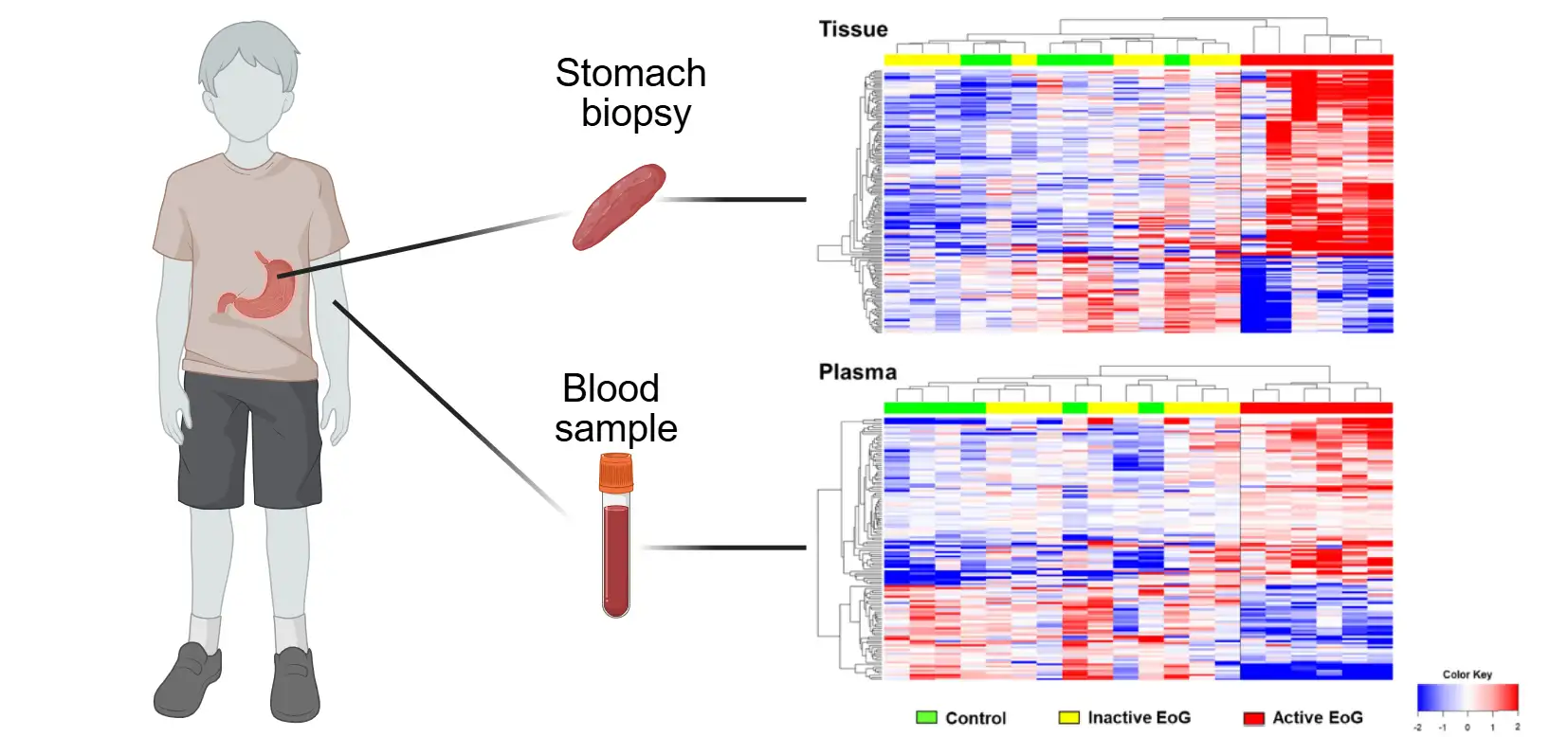
Researchers first performed high-depth proteomic analysis on paired gastric tissue and plasma from children with active EoG, inactive EoG and symptomatic controls without eosinophilic gastrointestinal disease. They detected 2,297 proteins in gastric tissue and 5,977 in plasma, then identified 25 proteins that were altered in both compartments.
These proteins represented several aspects of disease biology. Some were eosinophil granule proteins, while others were related to extracellular matrix remodeling, inflammation and cellular stress. Together, they formed a molecular signature that was also associated with clinical measures including blood and gastric tissue eosinophil counts.
The team then evaluated the candidate signature in an independent group of adults enrolled in the ELEMENT study. Paired serum samples were obtained during active disease and after elemental diet–induced clinical and histologic remission. Of the original 25 candidate proteins, 23 were detectable in serum samples from these participants, and the overall proteomic pattern shifted with treatment. Levels of several proteins changed as disease activity improved, further supporting the link between the blood-based protein signature and treatment response.
Looking Ahead
The researchers caution that the work is exploratory. The study included a relatively small number of participants, and additional research will be needed to determine how well the protein signature performs across larger and more diverse patient populations.
Still, the findings provide an important step toward developing blood-based approaches informed by what is happening directly in diseased stomach tissue. Notably, the protein signature captured more than eosinophil-related inflammation, reflecting structural and tissue-remodeling processes that may contribute to long-term disease activity.
“Our long-term goal is to reduce reliance on repeated invasive procedures while still understanding what is happening in the tissue,” says Shoda.
Future studies will help determine whether these molecular signals can complement biopsy-based assessments and improve how EoG activity and treatment response are monitored over time.
About the Study
The study reflects a broader team-science model at Cincinnati Children’s, including a collaboration with the Divisions of Gastroenterology, Hepatology and Nutrition and Allergy and Immunology and the Cincinnati Center for Eosinophilic Disorders (CCED). Molly Ahye led the work during her pediatric gastroenterology fellowship while working closely with senior investigators and collaborators, including Marc Rothenberg, MD, PhD, director of the CCED, and Nirmala Gonsalves, MD, whose ELEMENT cohort provided the longitudinal treatment-response samples. The project illustrates how fellowship research can be integrated into established clinical and translational programs.
External collaborators included experts from Northwestern University, the Kazusa DNA Research Institute, Chiba University, the University of Cincinnati, and the National Institute of Allergy and Infectious Diseases.
Funding for the study was supported by the Gastroenterology T32 Training Grant (T32 DK007727) and the Intramural Research Program of the National Institutes of Health (NIH).
Don’t Miss a Post:
- Subscribe to the Research Horizons Newsletter
- Follow Cincinnati Children’s Research Foundation on Bluesky, X and LinkedIn
| Original title: | A tissue-informed blood proteomic signature of eosinophilic gastritis activity |
| Published in: | The Journal of Allergy and Clinical Immunology |
| Publish date: | October 7, 2026 |
Research By

The Shoda Lab at Cincinnati Children’s is working to identify the key genes and genetic regulatory mechanisms that predispose patients to eosinophilic conditions, especially eosinophilic gastrointestinal diseases (EGID).
Latest Posts
About this blog
The Research Horizons blog features news and insights about the latest discoveries and innovations developed by the scientists of Cincinnati Children's. This blog does not provide medical advice, diagnosis, or treatment. Email researchnews@cchmc.org with questions or ideas.
Subscribe to Our Newsletter