Scientists Unlock Scalable Production of Human Gut Organoids with Functional Nerves
Research By: Holly Poling, PhD | Maxime Mahe, PhD | James Wells, PhD | Michael Helmrath, MD
Post Date: May 22, 2026 | Publish Date: May 22, 2026
“Confined culture system” developed at Cincinnati Children’s opens doors for faster, wider use of lab-grown human tissues in disease studies and drug development
Thanks to special 3D-printed scaffolding trays designed by experts at Cincinnati Children’s, researchers can now produce larger versions of functional human gut organoids twice as fast as previous methods—and these organoids grow their own nerve cells.
This improved technology could help accelerate production of human mini-organ tissues that are large enough to be useful in patching damage or restoring diminished functions of a person’s small intestine, stomach or colon. Such tissues also would be valuable for future disease studies and to more accurately evaluate organ damage risks linked to oral medications.
Details of the project were published May 22, 2026, in Nature Biomedical Engineering. The new production system was designed and tested by a team led by staff investigator Holly Poling, PhD, senior author Maxime Mahe, PhD, and a team of 17 other scientists from Cincinnati Children’s and Nantes Université in France.
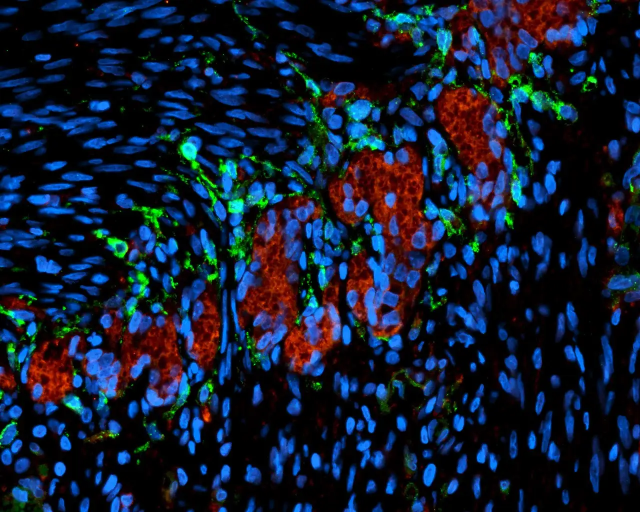
Using their new “confined culture system” (CCS), the team grew small intestine, colon, and stomach organoids from tiny spherical forms into centimeter-scale tubular forms nearly 10 times larger than previous methods. Also, unlike methods that require a complex effort to introduce nerve cells, these organoids develop a nervous system on their own.
“By reaching transplantation maturity twice as fast and developing their own functional nerves, these organoids demonstrate how engineering principles can drive biological innovation,” Poling says. “Our confined culture system is more than a production method; it’s a scalable, flexible platform for building complex human tissues.”
New production system prompts faster growth
Experts at Cincinnati Children’s Center for Stem Cell & Organoid Medicine (CuSTOM) have been leaders at making miniature versions of digestive system organs for more than 15 years, steadily improving the sophistication of the lab-grown tissues. More recently, the team has been working on methods to make enough customized tissue to transplant into patients to help patch organ damage or restore diminished specialized functions.
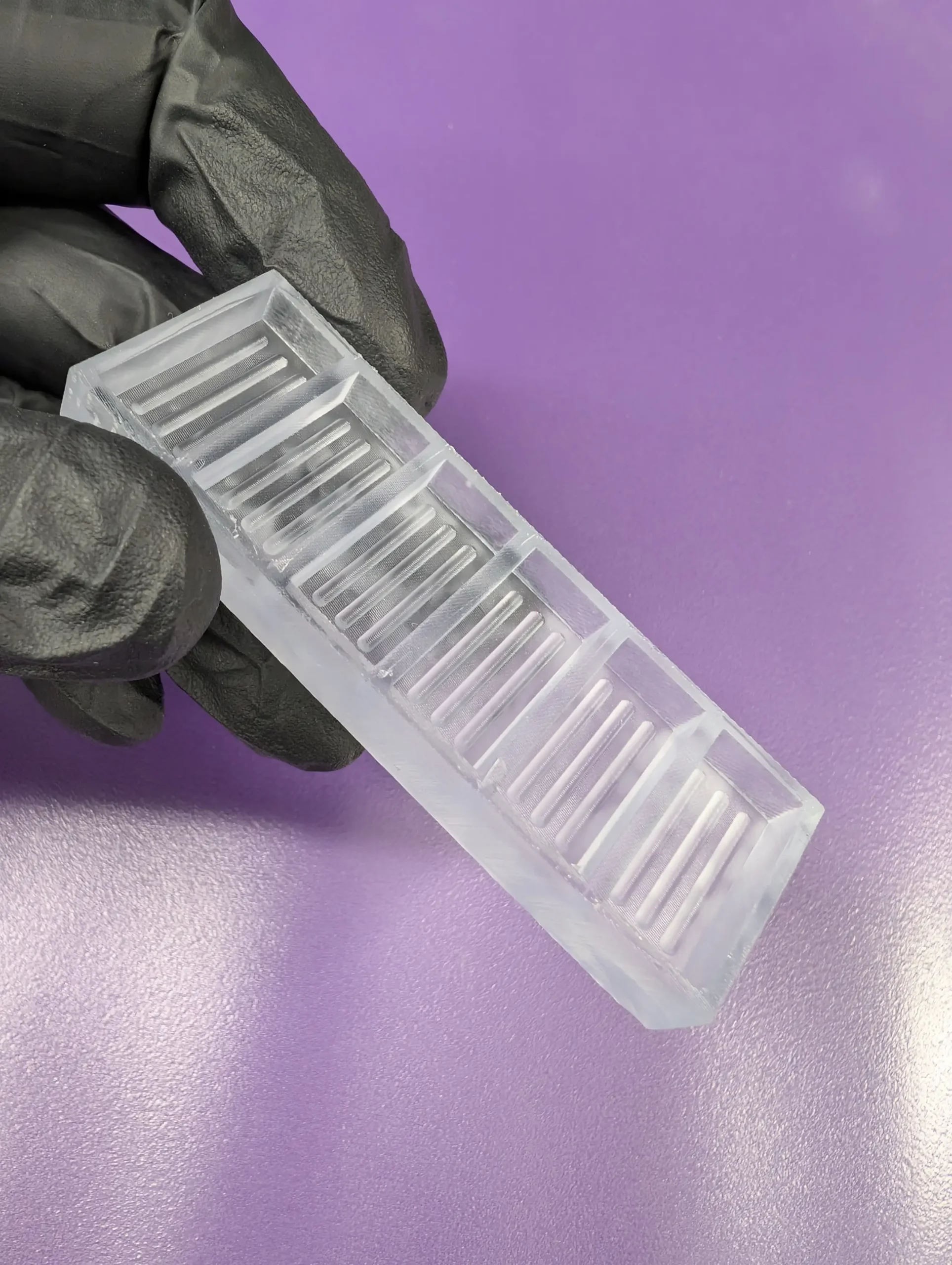
The new method uses 3D printing technology to make tray-like scaffolding molds from surgical resin, then filling the molds with degassed polydimethylsiloxane—a flexible rubber-like type of silicone.
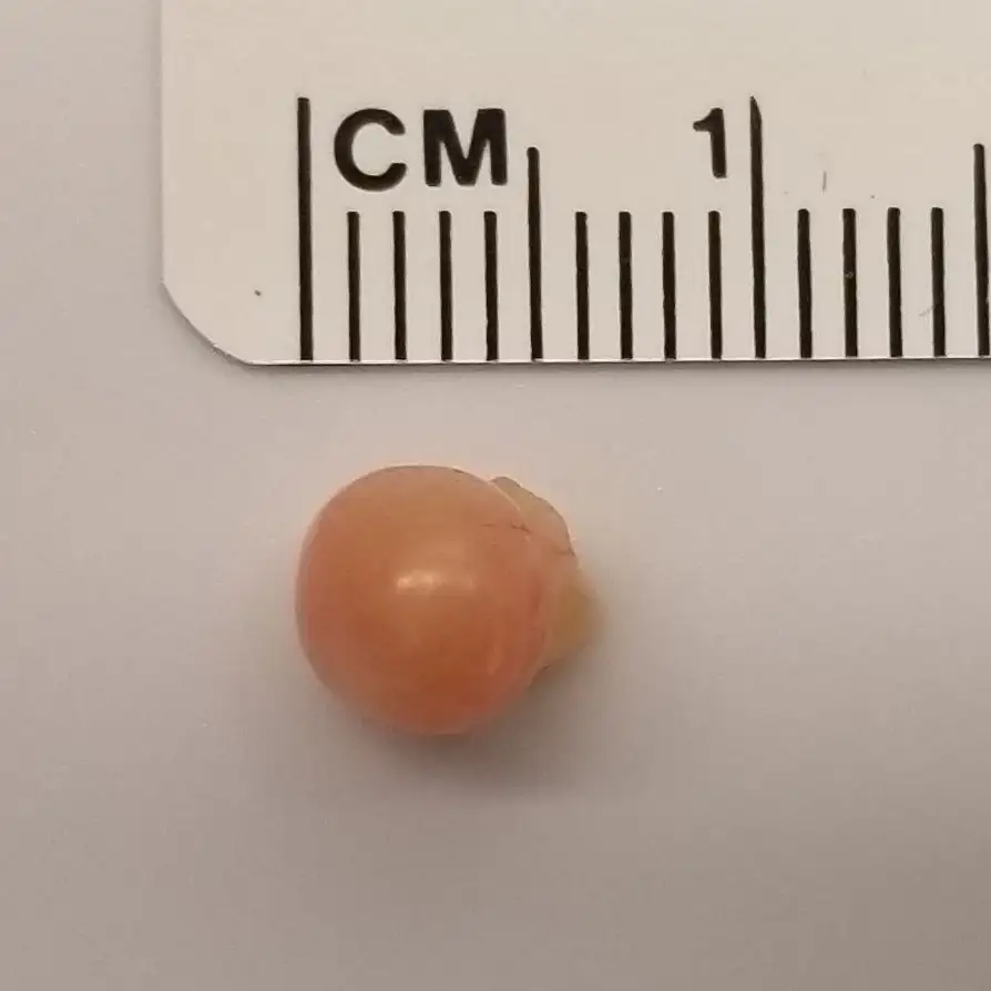
The new trays contain grooves designed to confine a collection of sphere-shaped organoids into a row, which encourages the spheroids to fuse together and mature. The fusions occur within a special mix of nutrients and other ingredients that support initial growth from induced pluripotent stem cells (iPSCs) into more complex organoids.
By day six, the discrete spheroids develop into unified constructs along the grooves of the trays. These are moved into another hydrogel medium for continued growth for another eight days.
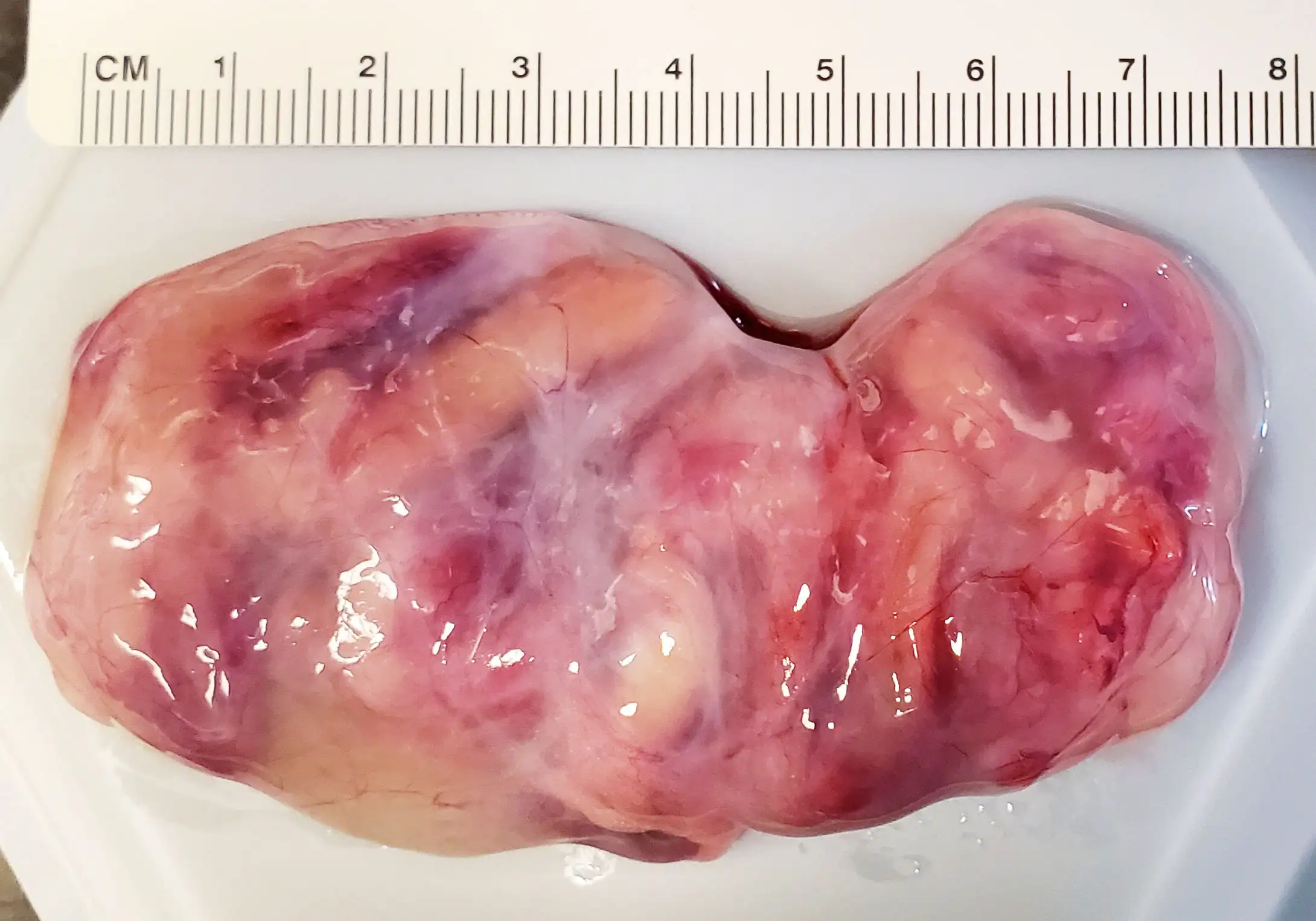
By day 14, the organoid constructs have produced all the cell types and structures that previously required 28 days to achieve. These tissues are then transplanted into rodents that are genetically modified to minimize rejection risk.
All of the transplanted tissues engrafted successfully in the rodents, the co-authors state. After growing in the rodents, the team produced as much as 8 cm of functioning small intestine tissue, compared to approximately 1 cm of tissue using previous protocols. Not only were the structures much larger than previous methods, but now their neuromuscular function was also similar to native human tissue, representing a major advance.
“We are now able not only to generate complex gastrointestinal organoids at scale, but also to guide their differentiation into functional tissues with integrated enteric neuronal networks,” Mahe says. “By leveraging a defined growth environment, the intrinsic self-organization capacity of the cells drives the formation of tissue structures that closely resemble the human gastrointestinal tract.”
Jim Wells, PhD, a study co-author and chief scientific director at CuSTOM says the new technology overcomes key barriers to scale and function in organoid research and biomanufacturing.
“This platform’s simplicity, reproducibility, and versatility make it accessible for widespread adoption,” Wells says. “In addition, the emergence of a self-organized nervous system within these organoids is particularly important for further studies of neurodevelopmental disorders.”
Another step closer to human clinical trials
Michael Helmrath, MD, a surgeon-scientist at Cincinnati Children’s who co-directs CuSTOM, has been working for more than a decade to develop intestine organoids sophisticated enough for transplantation in human patients.
In 2017, Helmrath and colleagues demonstrated how to combine neural crest cells with intestinal tissue cells in a layered process to make the first human organoids with nerve function. His team also showed how intestine organoids could be grown larger by implanting them in a mouse to provide a blood supply. Ever since, intestine organoids have been getting more sophisticated, including versions with immune cells in addition to the specialized organ cells and nerves.
Now the new process—involving rats instead of mice—produces even larger amounts of tissue.
“It is still not possible to grow complete, full-sized human organs in some sort of tank, but research like this has produced significant amounts of tissue that can be matched directly to individual patients,” Helmrath says. “We believe such tissues, once transplanted, would further grow and multiply as part of the patient’s own organ to restore functions.”
More research and development is needed before “CCS organoids” will be ready for human clinical trials, Helmrath says. But if successes continue, organoid medicine may allow more infants and children with dysfunctional organs to be treated without ever needing a full organ transplant.
About the study
In addition to Poling, Mahe, Wells and Helmrath, Cincinnati Children’s co-authors included Akaljot Singh, Garrett Fisher, Konrad Thorner, Praneet Chaturvedi, Kalpana Nattamai, Kalpana Srivastava, Matthew Batie, Nicole Brown, Taylor Hausfeld, Amy Pitstick, Riccardo Barrile, Christopher Mayhew, and Takanori Takebe. Three experts with Inserm and Nantes Université also were co-authors.
Funding sources for the research included the National Institute of Diabetes and Digestive and Kidney Diseases (U01 DK103117, P30 DK078392) and the Agence Nationale de la Recherche (ANR-17-CE14-0021, ANR-21-CE14-0017).
| Original title: | Large-scale and innervated functional human gut tissues for transplantation via transient spheroid confinement |
| Published in: | Nature Biomedical Engineering |
| Publish date: | May 22, 2026 |
Research By


Research in the Wells lab aims to uncover the molecular and cellular mechanisms by which gastrointestinal and endocrine organs form in the developing embryo.
As a pediatric surgeon scientist, Dr. Helmrath has established a large multidisciplinary team dedicated to clinical, translational, and basic science research focused on human diseases.

Latest Posts
About this blog
The Research Horizons blog features news and insights about the latest discoveries and innovations developed by the scientists of Cincinnati Children's. This blog does not provide medical advice, diagnosis, or treatment. Email researchnews@cchmc.org with questions or ideas.
Subscribe to Our Newsletter