

Improving Early Detection of Graft Rejection
Research By: Anthony Sabulski, MD
Post Date: September 30, 2026 | Publish Date: May 28, 2026

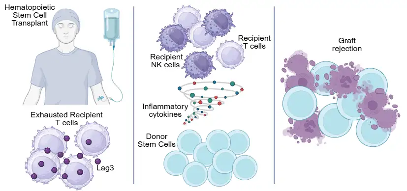
Researchers at Cincinnati Children’s identified blood-based markers that may help detect graft rejection earlier, giving clinicians a better opportunity to intervene before transplant failure progresses
Graft rejection after hematopoietic stem cell transplant (HSCT) remains one of the most serious and difficult complications in transplant care. Once it begins, it can progress quickly, often leaving few treatment options beyond repeat transplant. Clinicians can identify patients at higher risk, but there has been no reliable way to determine, early and precisely, which patients are most likely to need rapid intervention.
A study published May 28, 2026, in Blood Insights & Cellular Therapy helps define how graft rejection may be identified and managed earlier. Findings from this work were recognized with a Best Abstract Award at the 2024 Tandem Meetings of American Society for Transplantation and Cellular Therapy and the Center for International Blood and Marrow Transplant Research.
Researchers in the Division of Bone Marrow Transplantation and Immune Deficiency identified signals in the blood—such as CXCL11, CXCL10 and LAG3—that are linked to graft rejection and can appear before symptoms are fully recognized. The study also helps clarify how the immune system responds during rejection, offering direction for treatments aimed at controlling that process.
“We’re beginning to see clear signals in the blood that point to graft rejection before it fully develops,” says Tony Sabulski, MD, first author of the study. “That’s important because it gives us a chance to identify patients who may be heading toward rejection and consider treatment sooner, rather than waiting until the graft is already failing.”
Understanding Graft Rejection
Graft rejection occurs when the patient’s immune cells—particularly T cells and natural killer (NK) cells—recognize donor cells as foreign and begin to attack them. A key part of this response involves interferon, a signaling molecule produced by immune cells, including T cells, that activates inflammation.
Earlier studies have identified measurable signs of this activity. CXCL9, a protein produced in response to interferon, is elevated in patients with graft rejection. Other markers, including BAFF and indicators of complement activation, have also been linked to rejection. In addition, patterns such as higher and later fevers help distinguish graft rejection from other post-transplant complications.
Together, these findings highlight interferon as a central driver of graft rejection and support therapies that target this pathway. Clinical experience at Cincinnati Children’s, including studies using emapalumab, further supports this approach. However, identifying which patients are most at risk—and when to intervene—remains a key challenge.
Defining the Signals of Graft Rejection
To better understand graft rejection, the study compared patients who rejected their transplant with patients who had fevers but did not lose their graft.
They found a small set of proteins in the blood that were higher in patients with rejection. The strongest markers were CXCL11 and CXCL10, which are linked to immune activation, along with LAG3, a marker of T cell activity. Notably, some of these markers appeared early in the transplant course.
The study also looked at the immune cells involved at the time of rejection. There was a clear increase in patient immune cells, including natural killer (NK) cells and T cells.
In addition, the researchers identified signs of a specific type of cell damage—ferroptosis—in donor cells during rejection. This finding helps explain how donor cells may be eliminated.
“We’re not just identifying markers in the blood—we’re also seeing how those signals connect to the immune cells driving rejection and the process by which donor cells are lost,” says Sabulski.
Looking Ahead
This study helps shift graft rejection care from reacting to problems to identifying risk earlier and acting sooner.
Three areas stand out:
- Earlier identification of risk: Biomarkers such as CXCL11, CXCL10 and LAG3 may appear early—sometimes before clear symptoms—helping identify patients who need closer monitoring or earlier treatment.
- Stronger rationale for targeted therapies: The central role of interferon supports therapies designed to control this response, particularly in patients identified through biomarker monitoring.
- A more individualized approach to care: Combining risk factors with early biomarker signals may help tailor care to each patient and reduce the need for repeat transplant.
“As we build on these findings, the goal is to identify risk earlier and use that information to guide more targeted care, so we can reduce graft rejection and improve outcomes for patients undergoing transplant,” says Sabulski.
About the Study
Stella Davies, MD, was the senior and corresponding author of the study. Additional Cincinnati Children’s co-authors include Aaron Webster, Lucy Langenberg, Nathan Luebbering, Kasiani Myers, MD, Christopher Dandoy, MD, MSc, Diana Clark, Alana Snyder and Sonata Jodele, MD.
External co-authors include experts from Harvard Medical School and Beth Israel Deaconess Medical Center.
This study was funded by a Hyundai Hope on Wheels Young Investigator Award.
Don’t Miss a Post:
- Subscribe to the Research Horizons Newsletter
- Follow Cincinnati Children’s Research Foundation on Bluesky, X and LinkedIn
| Original title: | Plasma proteomics and single-cell RNA-seq identify cellular and soluble mechanisms of graft rejection after HSCT |
| Published in: | Blood Immunology & Cellular Therapy |
| Publish date: | May 28, 2026 |
Research By

Latest Posts

About this blog
The Research Horizons blog features news and insights about the latest discoveries and innovations developed by the scientists of Cincinnati Children's. This blog does not provide medical advice, diagnosis, or treatment. Email researchnews@cchmc.org with questions or ideas.
Subscribe to Our Newsletter