

Does Earlier Cleft Bone Grafting Help Teeth? Study Says No.
Research By: Jue Wang, DDS
Post Date: November 12, 2025 | Publish Date: Nov. 12, 2025

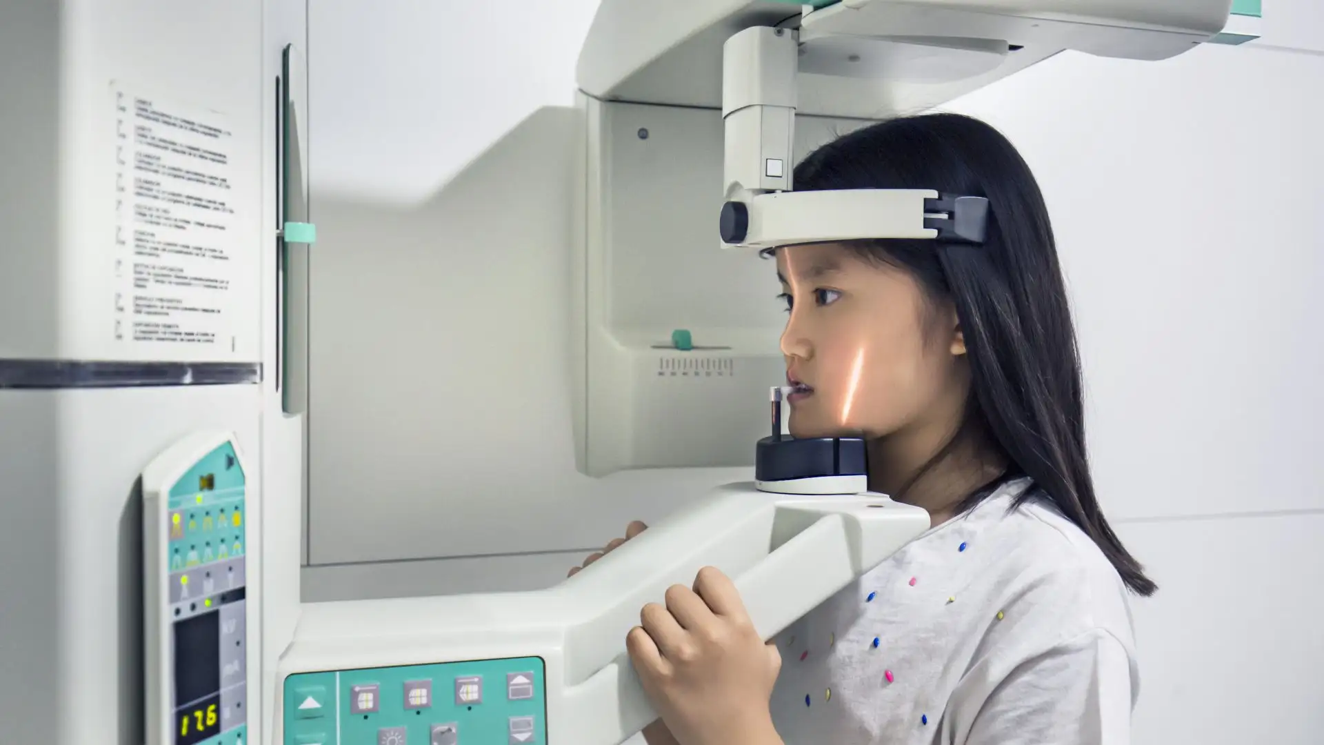
A Cincinnati Children’s study of 101 patients with cleft lip and palate found that a tooth’s location at the cleft margin, not the patient’s age at the time of alveolar bone grafting, affected incisor development
Key Takeaways
- Earlier bone grafting showed no advantage over later grafting for incisor development.
- Incisors bordering the cleft were more likely to form abnormally, regardless of the patient’s age at grafting.
- Later grafting leaves time for orthodontic preparation before surgery.
- Cone beam CT (CBCT) is a valuable way to evaluate tooth morphology and grafting outcomes.
Many children born with cleft lip and palate need alveolar bone grafting, a surgery that fills the gap in the upper gum line. The graft supports nearby teeth, helps permanent teeth erupt and stabilizes the upper jaw. Typically, most centers, including the Cleft and Craniofacial Center at Cincinnati Children’s, perform this grafting on patients between ages 9 and 12, before the canines erupt.
Some centers are moving the timeline earlier, to around age 6 or 7, hoping to protect the development of the incisors next to the cleft. So far, evidence for shifting the timeline has been limited. Previous studies have focused mostly on the bone graft itself.
A study at Cincinnati Children’s, published in The Cleft Palate Craniofacial Journal, looked at the teeth instead.
“A lot of papers look at whether the bone is thick enough or wide enough, but they never look at the teeth,” says corresponding author Jue Wang, DDS, PhD, an orthodontist in the Division of Pediatric Dentistry and Orthodontics. “From a dentist’s standpoint, I wanted to look at the tooth and see if early grafting really provides benefit, as others have proposed.”
Measuring the Teeth in 3D
Wang and her team retrospectively analyzed cone beam CT (CBCT) scans, a type of 3D X-ray, from 101 patients with cleft lip and palate who received treatment at Cincinnati Children’s. In the central and lateral incisors, they measured:
- Crown size
- Root length
- Root shape abnormalities
- Crown-to-root ratio
Then, they compared patients by age at the time of grafting: early (6–7 years), middle (8–9 years) and late (10 and older).
Location Is More Important Than Timing
They found that age at grafting was not associated with significant differences in crown size or root length or shape. However, position was. Central incisors bordering the cleft had narrower, shorter crowns and more root shape abnormalities than those farther away. Abnormal roots appeared in about 34% of upper right central incisors on the cleft margin versus 8% of those not on the margin, and in 49% versus 8% of upper left central incisors.
“We realized that maybe it’s not because we didn’t do the bone grafting early enough,” Wang says. “Maybe it’s just because of the location. The tooth doesn’t form very well, no matter if you put the bone in early or not.”
What the Findings Mean for Clinicians
The study’s findings do not support earlier grafting to improve incisor development, so clinicians considering graft timing can set tooth formation aside as a reason to graft sooner, Wang says. Later grafting also leaves time for orthodontic preparation before surgery, which can improve surgical access and arch alignment.
Other factors still matter. Some studies link early grafting to more bone at the graft site. Wang and her co-authors note that their single-center study was retrospective and lacked data on tooth eruption ages. Larger prospective studies could strengthen the findings.
Still, Wang says the Cincinnati Children’s cleft team will keep its current protocol.
“The results help us be more comfortable with our current protocol, knowing it’s the best evidence-based protocol we have for our patients,” she says.
Next Steps
Wang and her surgical colleagues are now comparing grafts made with the patient’s own bone (autograft) and donor bone (allograft). They are evaluating results from both surgical and dental perspectives, asking how much bone remains and whether it’s good enough to stabilize and guide the teeth.
Wang credits the multidisciplinary cleft team at Cincinnati Children’s, its large patient population, long-term follow-up and routine 3D imaging of graft outcomes for making this type of research possible.
About the Study
Co-authors include Jenna Panek, DMD, Jeffrey Lee, DDS, and Elizabeth Gosnell, DDS, from the Division of Pediatric Dentistry and Orthodontics; Qin Sun, MPH, from the Division of Biostatistics and Epidemiology; and John Jones from the University of Cincinnati College of Medicine.
Funding was supported by the Internal Division of Pediatric Dentistry and Orthodontics.
Don’t Miss a Post:
- Subscribe to the Research Horizons Newsletter
- Follow Cincinnati Children’s Research Foundation on Bluesky, X and LinkedIn
| Original title: | Effect of Timing of Secondary Alveolar Bone Grafting on Maxillary Incisor Development in Patients With Cleft Lip and Palate |
| Published in: | The Cleft Palate Craniofacial Journal |
| Publish date: | Nov. 12, 2025 |


Latest Posts

About this blog
The Research Horizons blog features news and insights about the latest discoveries and innovations developed by the scientists of Cincinnati Children's. This blog does not provide medical advice, diagnosis, or treatment. Email researchnews@cchmc.org with questions or ideas.
Subscribe to Our Newsletter