

Tiny Messengers May Help Explain How Bariatric Surgery Improves Metabolic Health
Research By: Takahisa Nakamura, PhD
Post Date: November 3, 2025 | Publish Date: September 10, 2025

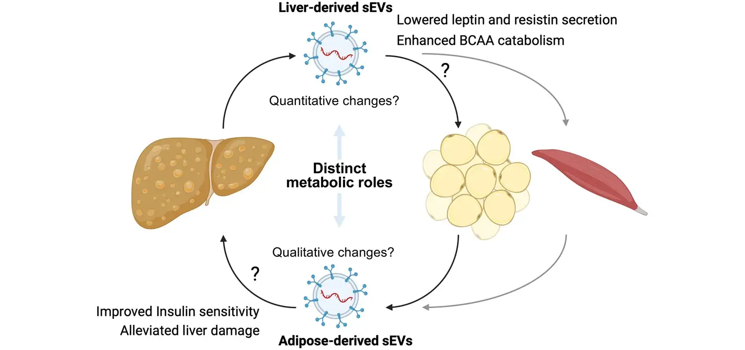
A Cincinnati Children’s study of adolescents undergoing bariatric surgery found that molecular signals exchanged between organs may contribute to metabolic improvements beyond weight loss alone
Bariatric surgery is one of the most effective treatments for severe obesity, yet many of its benefits occur far sooner than weight loss alone would predict. A Cincinnati Children’s study points to one possible explanation: tiny particles called extracellular vesicles that help organs communicate with each other.
Researchers led by Takahisa Nakamura, PhD, examined small extracellular vesicles (sEVs) in blood samples collected from 58 adolescents with severe obesity before and after vertical sleeve gastrectomy, the most common bariatric procedure performed in youth. These vesicles carry RNA and other molecular cargo throughout the body, providing a window into how different organs respond to surgery.
The team found that circulating sEVs carrying RNA signatures associated with the liver decreased significantly after surgery, while RNA signatures associated with adipose (fat) tissue remained relatively stable. Although both were associated with metabolic improvements, they appeared to be connected to different aspects of recovery.
Liver-enriched RNA cargo was most closely linked to broader metabolic changes. Lower levels of this cargo was associated with BMI and leptin levels six months after surgery, as well as decreases in branched-chain amino acids, molecules linked to insulin resistance and metabolic disease.
Fat tissue-enriched RNA cargo showed a different pattern. Changes in this cargo was associated with improvements in insulin sensitivity and markers of liver health, suggesting a potential role in how the body processes and responds to nutrients after surgery.
Together, the findings suggest that the liver and fat tissue may play distinct but complementary roles in recovery after bariatric surgery. The work also highlights extracellular vesicles as promising biomarkers that could help researchers measure and monitor metabolic health.
“Understanding how organs communicate after bariatric surgery could help us identify new opportunities to improve metabolic health, including potential approaches for patients who are unable or unwilling to undergo surgery,” says Nakamura.
About the Study
Ahlee Kim, Tsuyoshi Okura and Kwangmin Choi were the co-first authors of the study. Additional Cincinnati Children’s co-authors included members of the Divisions of Diabetes and Endocrinology, Experimental Hematology and Cancer Biology, Biostatistics and Epidemiology, General and Thoracic Surgery, and Developmental Biology.
External collaborators included experts from the University of Cincinnati, Children’s Hospital of Los Angeles, and Tottori University Faculty of Medicine.
Funding for the study was supported by the National Institute of Diabetes and Digestive and Kidney Diseases (R01DK123181, R01DK134646), Cincinnati Children’s Hospital Medical Center (Academic and Research Committee (ARC), and Japan Society for the Promotion of Science (19KK0402).
Don’t Miss a Post:
- Subscribe to the Research Horizons Newsletter
- Follow Cincinnati Children’s Research Foundation on Bluesky, X and LinkedIn
| Original title: | Exploring Organ-Specific Extracellular Vesicles in Metabolic Improvements Following Bariatric Surgery in Youth With Obesity |
| Published in: | Obesity |
| Publish date: | September 10, 2025 |
Research By


Latest Posts

About this blog
The Research Horizons blog features news and insights about the latest discoveries and innovations developed by the scientists of Cincinnati Children's. This blog does not provide medical advice, diagnosis, or treatment. Email researchnews@cchmc.org with questions or ideas.
Subscribe to Our Newsletter