In Utero Opioid Exposure Impacts Fetal Brain Size and Development
Research By: Usha Nagaraj, MD | Stephanie Merhar, MD, MS
Post Date: September 28, 2022 | Publish Date: Sept. 28, 2022
Radiology | Top Scientific Achievement


The opioid epidemic in the United States doesn’t discriminate—even infants are at risk.
Infants exposed to prenatal opioids are at increased risk of cognitive and behavioral problems during childhood. A case-control study led by first author Usha Nagaraj, MD, along with Stephanie Merhar, MD, MS, and others, looked at a series of fetal MRIs of subjects in the third trimester of pregnancy to determine the effects of opioid exposure.
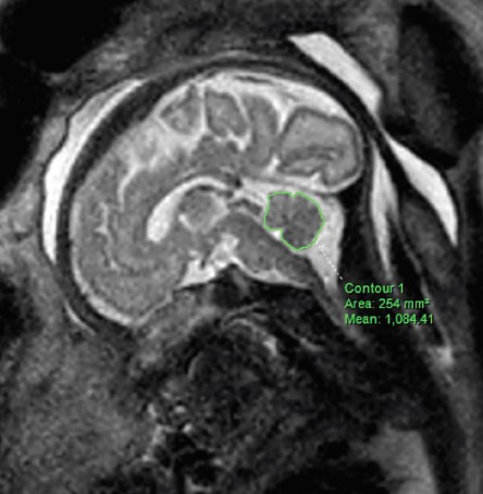
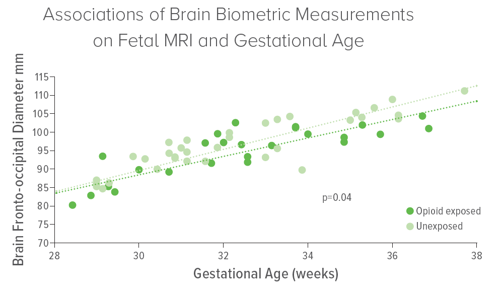
Fetuses were classified as opioid exposed or unexposed in utero, and 14 two-dimensional biometric measurements of the fetal brain were manually assessed and used to derive four indexes. Adjusting for gestational age, fetal sex and nicotine exposure, the researchers discovered that seven measurements were smaller in the opioid-exposed fetal brains, indicating smaller brain size and larger frontal occipital index. These measurements include cerebral frontal occipital diameter, bone biparietal diameter, brain biparietal diameter, corpus callosum length, vermis height, anteroposterior pons measurement and transverse cerebellar diameter.
Additionally, while differences in fetal motion, cervical length and deepest vertical pocket of amniotic fluid were statistically insignificant between the two groups, opioid-exposed fetuses showed higher frequencies of both breech position and increased volume of amniotic fluid. Nagaraj explains that the increased incidence of breech presentation may reflect neuromuscular dysfunction in utero and that while the increased amniotic fluid volume is of unclear etiology, the finding suggests an underlying physiologic aberration.
“The findings suggest that opioids may have an impact on development even prior to birth,” says Nagaraj. “And while it is unclear at this time what the smaller brain sizes seen in the opioid-exposed fetuses mean in regard to postnatal development, future studies in larger cohorts may help shed light on this.”

More 2023 Research Highlights
Chosen by the Department of Radiology
Wang J, Li H, Qu G, et al. Dynamic weighted hypergraph convolutional network for brain functional connectome analysis. Med Image Anal. 2023;87:102828. doi:10.1016/j.media.2023.102828
Abu-Ata N, Dillman JR, Rubin JM, et al. Ultrasound shear wave elastography in pediatric stricturing small bowel Crohn disease: correlation with histology and second harmonic imaging microscopy. Pediatr Radiol. 2023;53(1):34-45. doi:10.1007/s00247-022-05446-z
Dillman JR, Thapaliya S, Tkach JA, Trout AT. Quantification of Hepatic Steatosis by Ultrasound: Prospective Comparison With MRI Proton Density Fat Fraction as Reference Standard. AJR Am J Roentgenol. 2022;219(5):784-791. doi:10.2214/AJR.22.27878
Alves VPV, Dillman JR, Tkach JA, Bennett PS, Xanthakos SA, Trout AT. Comparison of Quantitative Liver US and MRI in Patients with Liver Disease. Radiology. 2022;304(3):660-669. doi:10.1148/radiol.212995
Somasundaram E, Castiglione JA, Brady SL, Trout AT. Defining Normal Ranges of Skeletal Muscle Area and Skeletal Muscle Index in Children on CT Using an Automated Deep Learning Pipeline: Implications for Sarcopenia Diagnosis. AJR Am J Roentgenol. 2022;219(2):326-336. doi:10.2214/AJR.21.27239
View more discoveries from 50 research divisions and areas
Return to the 2023 Research Annual Report main features
| Original title: | MRI Findings in Third-Trimester Opioid-Exposed Fetuses, With Focus on Brain Measurements: A Prospective Multicenter Case-Control Study |
| Published in: | American Journal of Roentgenology |
| Publish date: | Sept. 28, 2022 |
Research By


Merhar serves as attending neonatologist at the Perinatal Institute at Cincinnati Children’s. Her research focuses on how the newborn brain can bounce back from insults including brain injury and substance exposure.


