Exploring the Origins of Disease
Post Date: May 2, 2025 | Publish Date:
Developmental Origins of Health and Disease
Research Annual Report 2024

A never-ending quest for knowledge drives scientists at Cincinnati Children’s to collaborate across disciplines to shed new light on the developmental and childhood origins of disease.
Be they common conditions such as asthma or one of the thousands of rare diseases that can strike during childhood, basic research breakthroughs happening here can influence the direction of further studies for years to come. Cincinnati Children’s experts produce the early seeds of discovery that ultimately sprout into improved diagnostics and treatments not just for children, but for the benefit of people across the lifespan.
The following summaries highlight some of the far-reaching studies published in fiscal 2024 and featured on our Research Horizons Science Blog:

How ‘Pioneers’ Blaze the One Trail That Determines Cell Fate
A study exploring how pioneering cells determine the fate of other cells has significant implications for developmental biology. Understanding these processes could lead to advances in regenerative medicine and tissue engineering.
Findings were published online in Molecular Cell that reveal new details about how pioneer transcription factors do their important jobs.
“As we gain a more comprehensive understanding of the mechanisms underlying pioneer TF-mediated gene repression, it will greatly enhance the precise manipulation of cell fate in cellular programming and reprogramming,” says Makiko Iwafuchi, PhD, a member of the Division of Developmental Biology and the Center for Stem Cell & Organoid Medicine.
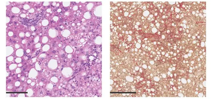
Profiling the Liver’s Frontline Defender
Cincinnati Children’s researchers have profiled the liver’s primary defensive cells, shedding light on their roles in maintaining health and combating disease. The paper, published in Nature Immunology, defines how the local environment in the liver and genetic background control gene expression in Kupffer cells.
“An important outcome of this study was finding that Kupffer cell transcription in different strains of mice was primarily controlled by non-cell autonomous trans effects of genetic variation rather than direct cis and cell autonomous trans effects,” says co-corresponding author Ty Troutman, PhD.
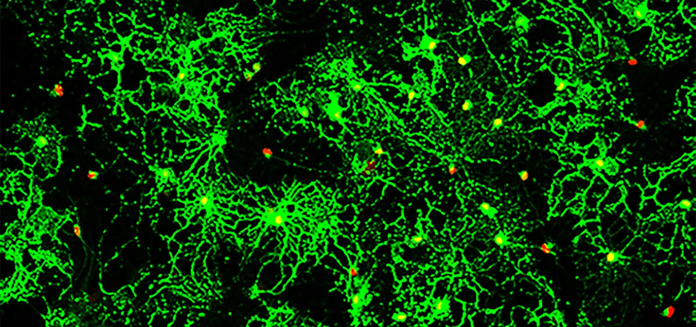
Small Molecule Shows Early-Stage Promise for Repairing Myelin Sheath Damage
When treated with a novel protein function inhibitor called ESI1, mice that mimic the symptoms of multiple sclerosis (MS) and lab-prepared human brain cells both demonstrated the ability to regenerate vital myelin coatings that protect healthy axon function.
This breakthrough, published in Cell, appears to overcome difficulties that have long frustrated previous attempts to reverse a form of nerve damage that robs people with MS of motor control and gradually blunts cognitive functions for many people as they age.
“Currently, there are no effective therapies to reverse myelin damage in devastating demyelinating diseases such as MS,” says corresponding author Q. Richard Lu, PhD. “These findings are significant as they offer new pathways for treatment that potentially shift the therapeutic focus from just managing symptoms to actively promoting repair and regeneration of myelin.”
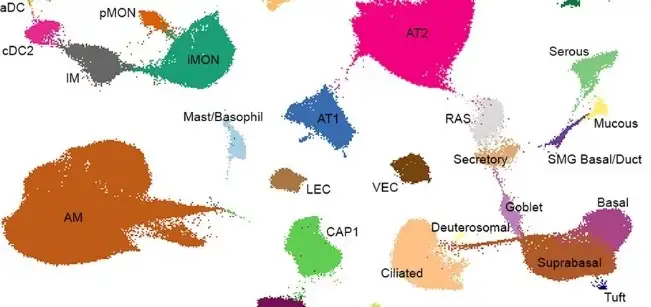
LungMAP Adds More Single-Cell References
The LungMAP project has expanded its single-cell references, providing researchers with an important batch of advanced single-cell reference data for deeper studies of lung development and disease. Details about the new data were published in Nature Communications.
“Our CellRefs, along with the developed analytic and web-based tools, are freely available to the pulmonary research community to facilitate hypothesis generation, research discovery, and identification of cell type alterations in disease conditions,” say corresponding authors Minzhe Guo, PhD, and Yan Xu, PhD.

Overactive Natural Killer Cells Linked to Asthma Progression
An unexpected hyperactive group of natural killer (NK) cells appears to play an important role in driving the “atopic march” from early eczema to increased risk of developing asthma later in childhood, according to a study in Science Immunology, led by Gurjit “Neeru” Khurana Hershey, MD, PhD, and colleagues.
“Our findings reshape the simple dogma of poor natural killer cell activity promoting eczema by demonstrating an unexpected wrinkle. An overactive population of natural killer cells in children with eczema may in fact worsen skin damage and provoke allergic sensitivity or development of asthma,” Hershey says.
This discovery could lead to new therapeutic strategies aimed at modulating the activity of these cells to manage asthma more effectively.
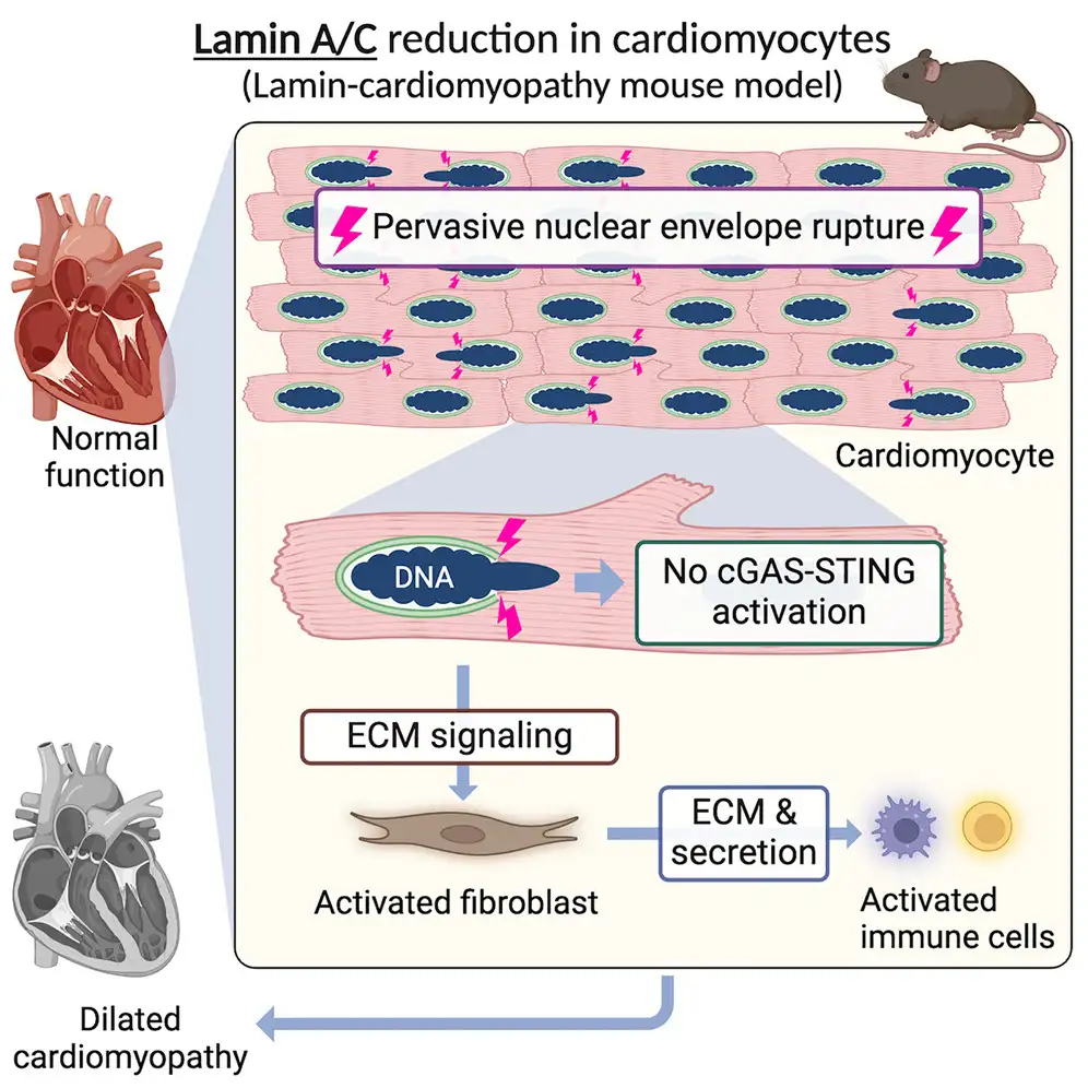
Study Challenges Current Thinking About Mechanisms of Dilated Cardiomyopathy
Nuclear envelope ruptures in heart muscle play a larger-than-expected role in driving dilated cardiomyopathy (DCM), a common form of heart disease, according to a study published online in Cell Reports.
The research focused on LMNA-related DCM, a genetically inherited form of the disease. These cases account for 5% to 13% of idiopathic dilated cardiomyopathies (not linked to alcohol abuse, ischemia, infections or other known factors). The findings suggest a potential early diagnostic marker and alternative targets for improved management of this serious condition.
“This work is the first to establish that nuclear envelope rupture occurs frequently in the disease model heart before the heart starts becoming weak,” says corresponding author Kohta Ikegami, PhD. “We found that almost 50% of heart muscle cells in the disease model heart have nuclear envelope ruptures compared to near zero found in normal hearts.”
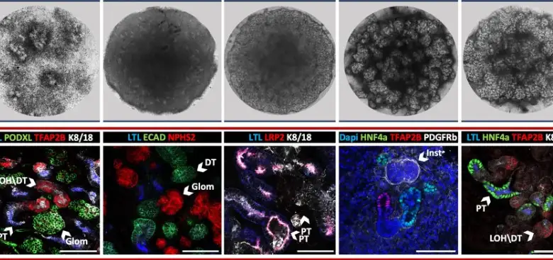
Organoid Technology Helps Reveal Possible Cause of Rare Fatal Kidney Disorder
Using rapidly advancing organoid technology, researchers led by Raphael (Rafi) Kopan, PhD, have uncovered key mechanisms that appear to drive a rare and fatal kidney disorder called autosomal recessive renal tubular dysgenesis (AR-RTD).
The work, published in Nature Communications, reveals that mutations in two genes can combine to delay kidney vascularization, thus starving renal tubules during a crucial point in their development. One mutation, occurring in angiotensin converting enzyme (ACE), blocks production of a molecular signal called angiotensin II (AngII). Another occurs in the gene AngII receptor type 1 (AGTR1), which prevents cells from “hearing” the AngII signal.
“The research suggests that a more promising approach to prevent AR-RTD would be to identify the critical nutrients that specifically support proximal tubule growth, and provide them as a supplement,” Kopan says.
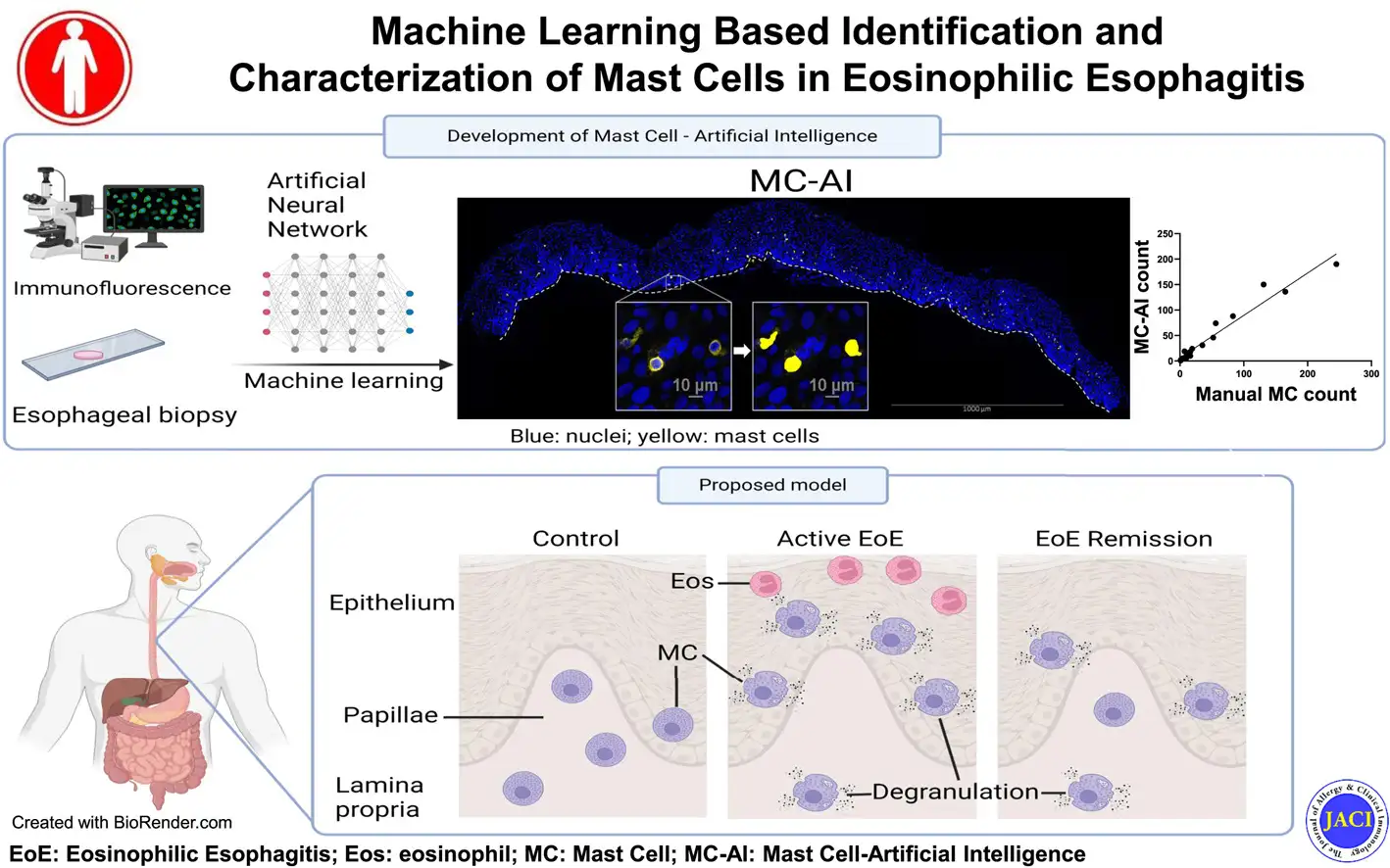
Artificial Intelligence Assists Expanded Hunt for Cells Driving Eosinophilic Esophagitis
A research team led by first author Simin Zhang, MD, developed a novel machine-learning protocol that helped identify a distinct population of homeostatic esophageal papillae mast cells that may contribute to the rare food allergy eosinophilic esophagitis.
The team named the new protocol Mast Cell-Artificial Intelligence (MC-AI) and shared details in the Journal of Allergy and Clinical Immunology.
“Mast cells are implicated in many diseases, including eosinophilic esophagitis, anaphylaxis, and even cancers. MC-AI can therefore be broadly applied to better understand the characteristics of this tissue-resident cell,” Zhang says.
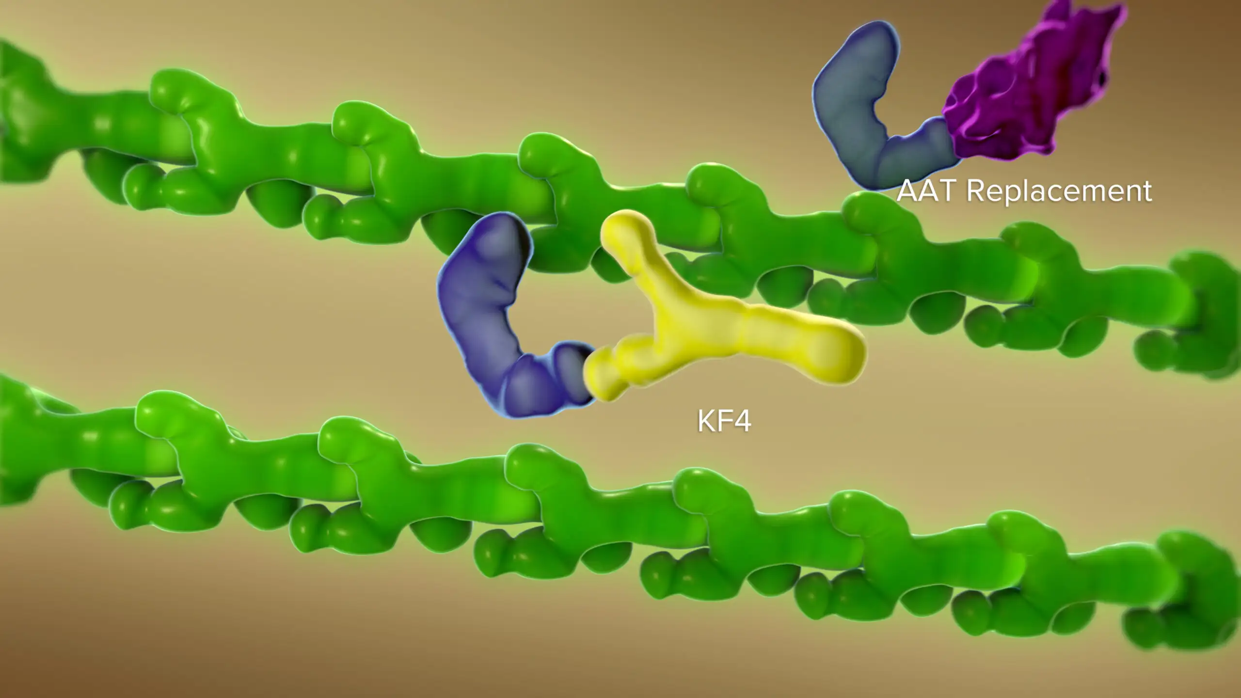
CELA-1 Antibody Shows Therapeutic Potential for COPD and Emphysema
In mice, a novel antibody that blocks chymotrypsin-like elastase 1 (CELA1) successfully reduced the lung damage caused by diseases such as age-related emphysema and chronic obstructive pulmonary disease (COPD), according to a study published in JCI Insight.
When CELA1 is introduced to adult lung specimens under physical stretch, it actively bonds to the lung tissue, which enhances the remodeling of elastase activity, a key disease process in lung disorders.
“By using a novel antibody to block CELA1, we observed a protective decrease in elastin remodeling. We were able to duplicate these protective effects in studies involving three types of chronic obstructive pulmonary
disease,” says Brian Varisco, MD, previously with the Division of Critical Care Medicine, and now with the University of Arkansas.
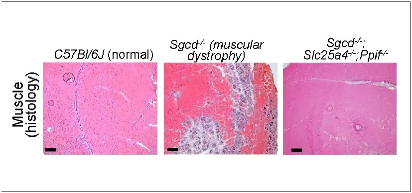
Mitochondria Pore Emerges as Potential Key to Managing Muscular Dystrophies
A study published in Science Advances reports an entirely new approach to preventing the muscle-wasting symptoms of muscular dystrophies. The research focuses on the role played by mitochondria, the tiny organelle
within our cells that processes nutrients into the energy cells need to survive.
“We have isolated the primary disease-causing component of muscular dystrophy to the mitochondrial permeability pore,” says Jeffery Molkentin, PhD. “If we prevent this pore from functioning, dystrophic disease in the mouse models we studied almost completely vanishes. We see the protection lasting past one year of life in the mouse, which translates to about 40 years of life for a human.”
Explore the 2024 Research Annual Report


Latest Posts


About this blog
The Research Horizons blog features news and insights about the latest discoveries and innovations developed by the scientists of Cincinnati Children's. This blog does not provide medical advice, diagnosis, or treatment. Email researchnews@cchmc.org with questions or ideas.
Subscribe to Our Newsletter