Organoid Revolution Happening Here
Research By: James Wells | Michael Helmrath, MD
Post Date: June 27, 2019 | Publish Date: January 2015
Top Research Achievements 2010-2015
Since 2010, scientists at Cincinnati Children’s have produced more than 7,000 peer-reviewed studies with far-reaching impact across pediatric medicine. From this impressive body of work, six advances stand out as Cincinnati Children’s premier scientific achievements of 2010-15.
Be they test platforms to evaluate new medications or the future of regenerative medicine, the tiny intestine and stomach organoids developed at Cincinnati Children’s by James Wells, PhD and colleagues have only just begun to demonstrate their potential to transform medicine.
In December 2010, a paper posted online in Nature revealed that Wells’ team had created functional, three-dimensional intestinal tissue from a combination of human embryonic stem cells (hESCs) and induced pluripotent stem cells (iPSCs). With this breakthrough, Cincinnati Children’s joined an elite class of medical centers making dramatic progress at growing miniature human brains, kidneys, pancreata, and other complex tissues in laboratory settings.
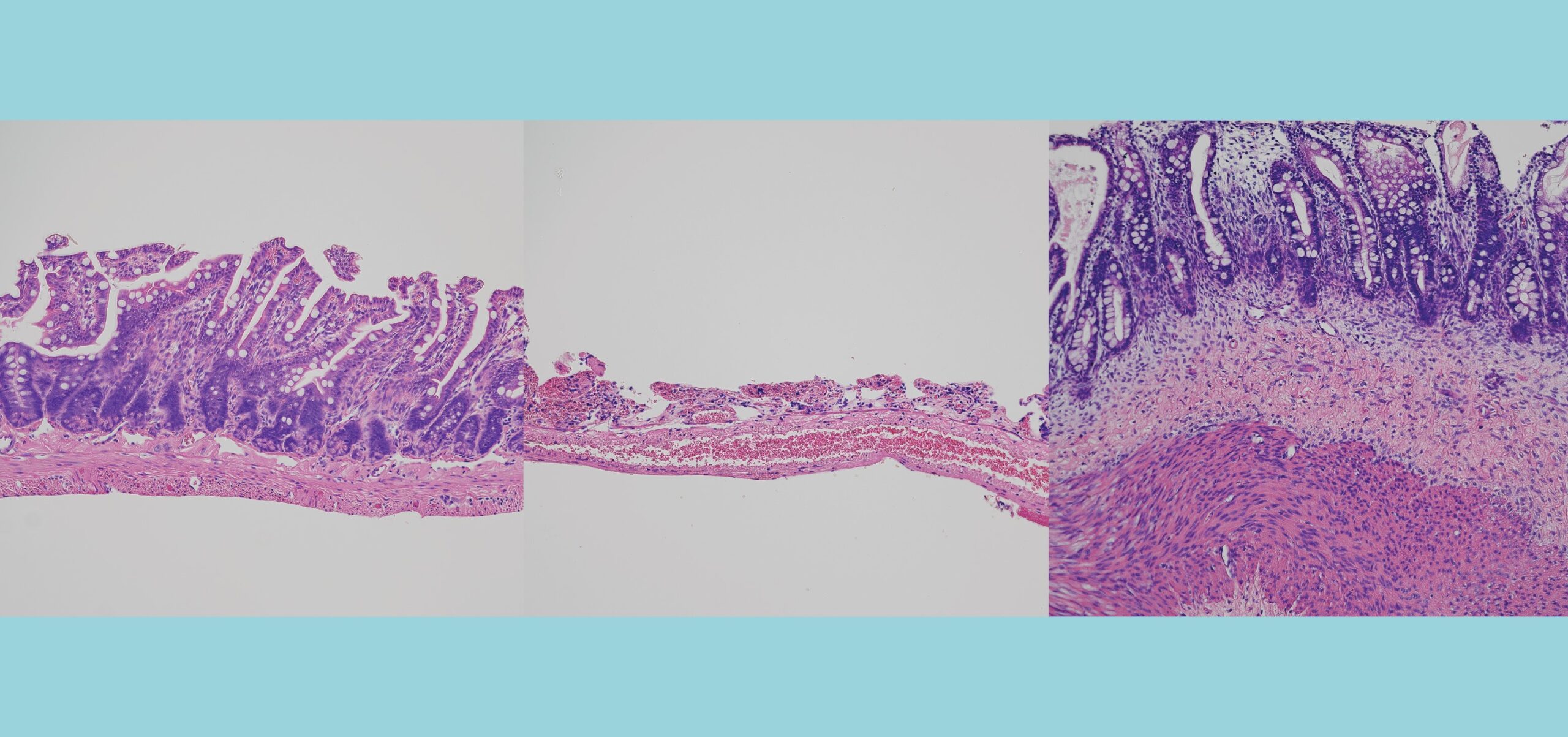
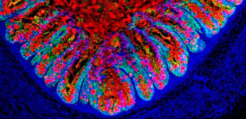
In a series of cell manipulations, Wells’ team coaxed stem cells to transform from definitive endoderm into hindgut progenitor cells, and ultimately into organoids containing all the major intestinal cell types – including enterocytes, goblet, Paneth and enteroendocrine cells. The tissue demonstrated absorptive and secretory functions and even began forming its own intestine-specific stem cells.
At this point, Wells’ team had an organ-on-a-chip, a research tool potentially capable of supplanting mice as a human-based model for studying disease. The mini-intestine shows promise as a model to further study necrotizing enterocolitis, inflammatory bowel disease, short bowel syndrome and more.
But the team did not stop here. In a study published online Oct. 19, 2014, in Nature Medicine, Wells collaborated with Michael Helmrath, MD, MS,, to demonstrate that intestinal organoids can grow into fully mature human tissue once grafted to a mouse kidney to provide a blood supply. This tissue included muscle layers and a self-renewing mucosal lining. The success was an encouraging sign that stem cell-derived organoids might be able to grow on their own once transplanted into the human body.
The very same month, Wells and graduate student Kyle McCracken published another paper in Nature announcing success at forming a mini-stomach. This “gastric organoid” specifically resembled the antrum, the portion of the stomach that connects to the intestine. Importantly, the organoids can harbor gut bacteria, which makes them immediately useful in research related to H. pylori, the bacterium that causes stomach ulcers.
Further organoid development continues at a rapid pace. Wells and colleagues are working on growing the fundus, the acid-secreting portion of the stomach and several other projects. An exciting aspect of the stomach project is that the organoids were developed from functional anterior foregut spheroids, which also serve as the developmental root of the pancreas and the lung.
In fact, Jason Spence, PhD, one of the colleagues who worked with Wells on the intestine organoid project, has carried on the work at the University of Michigan. In March 2015, Spence reported success at developing a lung organoid with structures resembling bronchi and alveoli that survived in the laboratory for 100 days.
Learn more about organoid research at Cincinnati Children’s
(This story originally appeared in the 2015 Research Annual Report)
Research By