Novel Smart Patch for Fetoscopic Myelomeningocele Repair
Post Date: February 16, 2021 | Publish Date:
Researchers at Cincinnati Children’s and the University of Cincinnati have developed a “smart patch” that could help significantly improve outcomes for babies with myelomeningocele (MMC).
The patch is designed for use during minimally invasive fetoscopic MMC repair and addresses the shortcomings associated with mesh-like patches currently in use.
Now, thanks to a $2.3 million grant from the National Institutes of Health (NIH), the research team has launched a study to further assess the patch’s efficacies in protecting the spinal cord. Principal investigators include Jose Peiro, MD, PhD, a fetal and pediatric surgeon at Cincinnati Children’s, and James Lin, PhD, a biomedical engineer at UC.
Cincinnati Children’s has been performing prenatal closure of MMC defects since 2011, when the Management of Myelomeningocele Study (MOMS) demonstrated the efficacy of prenatal surgery for children with this condition.
In 2016, surgeons at Cincinnati Children’s began doing fetoscopic MMC repairs, which have been shown to decrease morbidity in pregnant mothers and offer no inferior efficacy compared to open surgery. Fetoscopic MMC repair also maintains the neonatal patient benefits, such reverting the Chiari malformation, preserving motor function, and reducing hydrocephalus and the need for postnatal ventricular-peritoneal shunting.
A Shift to Fetoscopic Repair
When prenatal MMC repair is indicated, the fetal care team gives parents the option of an open or fetoscopic procedure. About 90% choose the fetoscopic technique.
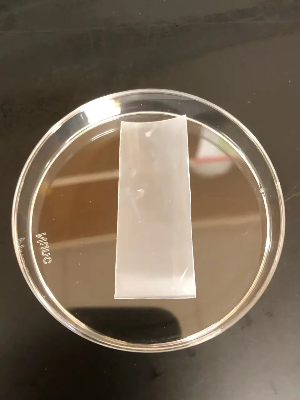
Currently surgeons use collagen patches to protect the spinal cord. Unfortunately, deploying these patches through small trocar ports and unfolding the patches for defect coverage can be cumbersome and prolong the duration of surgery. In addition, extensive suturing often is needed to create a watertight seal.
“We began looking for an alternative patch about four years ago, and when we couldn’t find an ideal one in the market, we decided to design it ourselves,” says Peiro, the director of endoscopic fetal surgery at Cincinnati Children’s Fetal Care Center. “Our team created a blend of two polymers — polylactic acid (PLA) and polycaprolactone (PCL). The material can be coiled and delivered through the cannula. Once inside the womb, it flattens against the spinal cord defect via body temperature activation, without any manipulation. The patch holds its shape and is water-tight, biocompatible and biodegradable.”
The Possibility of New Treatment Avenues

Peiro, Lin and their research team have tested the new smart patch in vitro, in vivo in rats, and in utero using neural progenitor cells. The new study is using an ovine model to assess the new patch in reducing surgery time, protecting the exposed spinal cord, and enhancing wound closure without tethering the spinal cord.
The team also will evaluate how the patch carries drugs such as growth factors and stem cells, which could open new treatment avenues.
Before joining Cincinnati Children’s in 2013, Peiro pioneered a fetal surgery program in Barcelona. He was one of the world’s first pediatric surgeons to perform fetoscopic MMC repair.
“This patch could represent an exciting improvement in fetoscopic MMC repair, which is already leading to promising outcomes in babies and reduced risks for mothers,” he says. “We hope to have it available in the clinical setting soon so that many patients can benefit.”
For more information, contact Jose Peiro at Jose.Peiro@cchmc.org.
Latest Posts


About this blog
The Research Horizons blog features news and insights about the latest discoveries and innovations developed by the scientists of Cincinnati Children's. This blog does not provide medical advice, diagnosis, or treatment. Email researchnews@cchmc.org with questions or ideas.
Subscribe to Our Newsletter