‘Myoatlas’ Reveals Some Muscle Fiber Nuclei Play Special Roles
Research By: Michael Petrany | Douglas Millay, PhD
Post Date: December 11, 2020 | Publish Date: Dec. 12, 2020

Scientists studying muscle development have established that muscle fibers form by fusing multiple myocytes into a cell that contains numerous nuclei working within a shared cytoplasm.
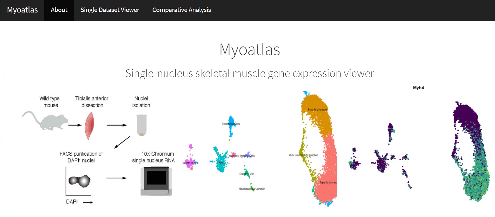
Now, data from a novel single-nuclei analysis shows that some of the nuclei within the fibers perform distinct tasks from the others, according to findings published Dec. 12, 2020, in Nature Communications.
The study, led by experts at Cincinnati Children’s, examined nuclei from mouse skeletal muscle across the lifespan. The researchers found distinct myonuclear populations emerging in postnatal development as well as aging muscle.
“Our datasets also provided a platform for discovery of genes associated with rare specialized regions of the muscle cell, including markers of the myotendinous junction and functionally validated factors expressed at the neuromuscular junction,” the co-authors wrote.
“This data gives an unprecedented glimpse into rare nuclei and expression patterns that help control skeletal muscle biology,” says senior author, Douglas Millay, PhD.
Cincinnati Children’s co-authors included first author Michael Petrany, Casey Swoboda, Kashish Chetal, Xiaoting Chen, Matthew Weirauch, and Nathan Salomonis.
The team also generated a publicly available website for this data: https://research.cchmc.org/myoatlas/
Read about more research in Nature Communications from Millay and colleagues
| Original title: | Single-nucleus RNA-seq identifies transcriptional heterogeneity in multinucleated skeletal myofibers |
| Published in: | Nature Communications |
| Publish date: | Dec. 12, 2020 |
Research By


