Novel Implant Promotes Osteochondral Tissue Regeneration
Research By: Patrick Whitlock, MD, PhD
Post Date: November 5, 2024 | Publish Date: August 2024
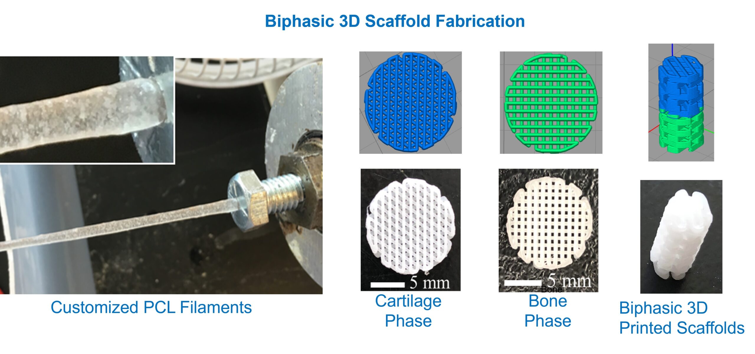
Study suggests a novel, 3D-printed scaffold may be a game-changing alternative to today’s limited treatment options
A 3D-printed implant developed by researchers from Cincinnati Children’s and the University of Cincinnati (UC) may lead to better treatments for large osteochondral defects.
Each customized implant, or scaffold, is made of synthetic and biological materials—including spherical microscopic particles (microspheres) containing decellularized human bone and cartilage tissue.
The research team recently investigated whether this biomimetic scaffold could regenerate functional osteochondral tissue in pre-clinical models. Their results suggest the scaffold may be an effective alternative to current treatment approaches, including the osteochondral autograft transfer system (OATS).
Current Treatments Associated with Significant Limitations
Due in part to the avascular nature of articular cartilage, large osteochondral defects—especially those greater than 2.5 cm in diameter—are difficult to treat. As a result, children, teens and young adults with these injuries have a much higher risk of developing early osteoarthritis.
“Although we’ve made progress within the field of cartilage regeneration and repair, we still have limited success treating large osteochondral defects,” says orthopaedic surgeon Patrick Whitlock, MD, PhD, co-director of Cincinnati Children’s Hip Preservation Program and the study’s principal investigator. “Current tissue engineering and regenerative medicine approaches are associated with poor remodeling and integration, and declining mechanical and biologic properties. It also can be challenging to find available donor tissue for patients who might benefit from an osteochondral allograft.”
A Unique Combination of Materials and Manufacturing
To find a better solution for large osteochondral defect repair, Whitlock and some of his colleagues teamed up with researchers from the UC departments of Biomedical Engineering and Orthopaedic Surgery.
Together, they developed (and patented) a novel process for 3D printing using microspheres containing decellularized cartilage or bone matrix. These microspheres slowly release proteins and growth factors that help promote osteochondral tissue regeneration. They also remain functional when embedded within a specific polymer material used in 3D printing.
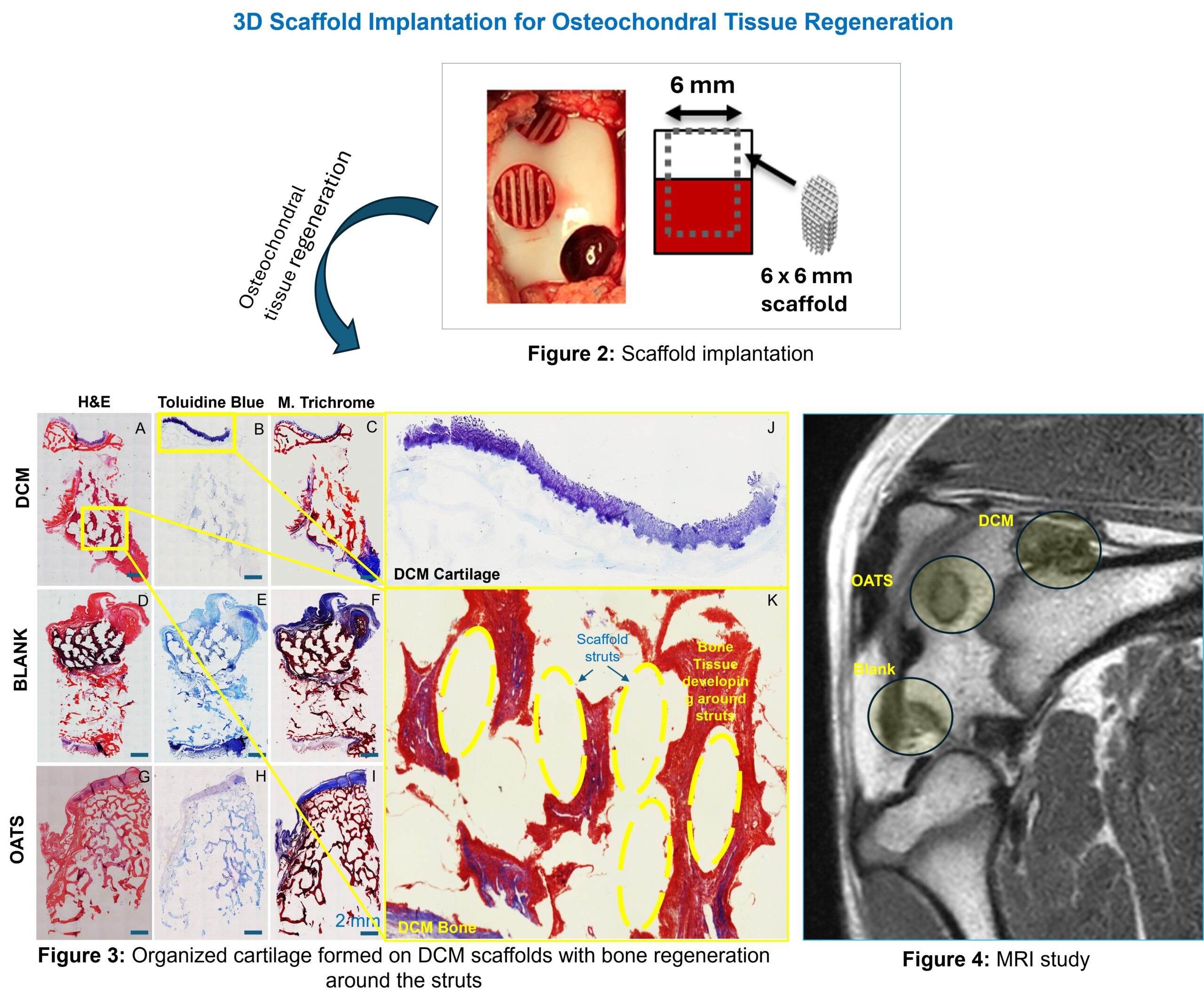
“Through our previous research, we figured out how to transform this novel material into three-dimensional scaffolds matched to the precise size and shape of individual osteochondral defects,” Whitlock says. “Then, in our most recent study, we assessed whether the scaffolds would induce osteochondral regeneration when integrated with live tissue.”
The Latest Research Yields Promising Results
The researchers implanted their biomimetic scaffolds in pre-clinical models with distal femoral osteochondral defects. For comparison, they also included negative controls (implanting scaffolds without decellularized human tissue) and positive controls (performing OATS autografts).
Whitlock says six months after implantation, the biomimetic scaffolds significantly outperformed their expectations.
“We found progressive, spatially oriented regeneration of osteochondral-like tissue that was at least comparable to—and in some cases, indistinguishable from—OATS autograft tissue,” he explains. “And our investigational scaffolds induced expression of certain bone and cartilage genes responsible for osteochondral tissue regeneration and maintenance.”
The team also found evidence of excellent scaffold integration with the host tissue.
“Collectively, the results suggest we’re on the right path with our research,” Whitlock says. “We’ll continue studying this scaffold in even larger osteochondral defects and in targeted pathologies, such as avascular necrosis of the femoral head. Hopefully, our findings will pave the way for treatments that restore normal joint function in patients with large osteochondral defects.”
| Original title: | Fabrication of a Biomimetic 3D Printed Scaffold for the Treatment of Large Osteochondral Defects in an Adolescent Porcine Model: Outcomes at 6 Months |
| Published in: | Journal of POSNA |
| Publish date: | August 2024 |
Research By
As a surgeon scientist, I have worked with our team to develop a multidisciplinary effort to advance the development, characterization and translation of naturally derived biomaterials to treat orthopaedic injuries and pathologies in children, adolescents and young adults.


